|

|
Disclaimer - While every effort has been made to ensure that the information that follows is accurate, MAF and HFRI do not accept liability for error of fact or opinion nor for the consequences of any decision based on this information.
© 1992 Ministry of Agriculture and Fisheries and the Horticulture and Food Research Institute. All rights reserved.
American foulbrood disease is caused by the bacterium Bacillus larvae. The disease is found world-wide, and is highly destructive to honey bee colonies. B. larvae is a spore-forming bacteria. The transfer of these spores spreads American foulbrood infection from colony to colony. B. larvae spores are very resistant to heat and chemical disinfectants. They can also withstand desiccation for at least 35 years.
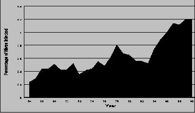
B. larvae was initially recorded in New Zealand in 1877, thirty-eight years after honey bees were first introduced. Today the incidence of B. larvae in New Zealand is reported to be approximately 1.2% of managed colonies and 7.0% of apiaries per year. The proportion of infected colonies is not high by world standards. However, the number of colonies and apiaries infected each year has increased over the last 40 years, with the sharpest increase occurring in the last 5 years (see graph).

The control of B. larvae is a major cost to the New Zealand beekeeping industry. The cost of the disease can be divided into two parts: the cost of the colonies that have to be destroyed, and the cost of inspecting colonies.
The total yearly cost of having to destroy colonies infected with B. larvae is almost impossible to determine. Besides the hives themselves the cost should include replacement colonies and equipment, transport, labour and lost production. The value of these components is likely to vary greatly depending on the individual beekeeping operation. However, at an estimated replacement cost of $150 per hive, the direct cost of American foulbrood to the New Zealand beekeeping industry was $560,000 for the 1990 - 1991 production year.
The cost of inspecting colonies for disease control is higher still, even though much of the inspection takes place while beekeepers are performing other tasks in the apiary. Industry estimates (labour only) range from $1,250,000 to $2,000,000 per year. The Ministry of Agriculture and Fisheries makes a further inspection input of about $135,000 per year.
The current control strategy in New Zealand hinges on the identification of colonies infected with B. larvae and their destruction by burning. Inspections are usually carried out in the spring because beekeepers have a statutory requirement to inspect their colonies and make a disease report to MAF by the 7th of December each year.
Many beekeepers also carry out further inspections during the year. Such inspections are often targeted for times when an undetected colony infected with B. larvae might result in the contamination of additional colonies ( e.g. when honey is being harvested or when hive parts are being exchanged).
No inspection method can detect the presence of B. larvae one hundred percent of the time (65% is the best detection rate so far achieved using visual inspection techniques). Inspecting anything less than all the brood frames in a hive decreases the chance of an American foulbrood infection being detected. And even checking every brood comb will not pick up very recent infections because diseased larvae may not become discoloured until 10 - 15 days after they have been infected. There is also the possibility that honey bee colonies may have "inapparent" B. larvae infections (that is, infections that do not exhibit outward symptoms).
Laboratory tests provide a means of detecting B. larvae infections which for one reason or another are not found by the beekeeper in the field. This manual describes a simple laboratory method for testing samples of adult bees and other material for the presence of B. larvae spores so that diseased colonies can be positively identified. The method consists of infecting bacteriological plates with test material, growing out bacterial cultures, and identifying the bacteria that result. The method is a simple, straight-forward test which can be carried out at home using readily available materials.
(While the method outlined in this manual is adequate for the purposes described, a variety of more sensitive and efficient tests exist which are more suited to fully equipped laboratories.)
The method described in this manual can be used to test a variety of material, including adult bees, larvae, pollen, and honey. However, two basic rules should be followed:
The bacterial culture method will work on older material, but there is a much greater chance of contamination affecting the results.
Sample Location in the Hive - take the bee sample from a brood comb. Do not shake any bees off first. Tests have shown that these bees are the ones most likely to be contaminated with B. larvae (see table).
(table)| Caste/Area | No. Bees Tested | No. Bees Testing Positive | Avg. B. Larvae Colonies/Bee |
| Workers On brood before shaking |
20 | 20 | 58.0 |
| Workers On brood after shaking |
20 | 16 | 16.0 |
| Workers On honey stores |
20 | 15 | 0.3 |
| Workers Leaving the colony |
10 | 4 | 6.1 |
| Workers Entering the colony |
10 | 2 | 0.6 |
| Drones | 20 | 13 | 0.2 |
Type of Container - the bees can be collected in any container provided that it is clean and has been sterilized if it has previously been in contact with bees or bee products. Recommended containers include plastic or glass jars and even plastic bags.
Number of Bees in the Sample - unless you have a good reason for doing otherwise, take at least 30 bees from the hive. If you are taking a composite sample (see "Composite Samples" in Preparing Samples below), take at least 60 bees from the hive.
How to Take the Sample - prop the comb to be sampled up against the side of a hive. Holding the container in one hand and the lid in the other, scrape the container rapidly up the comb, capturing the adhering bees. Before the bees can fly out, quickly put the lid back on the container. If a plastic bag is used, hold the bag open with both hands, scrape up the comb, and then twist the bag closed. Don't worry about killing the bees. They will die on their own in the sample container.
Hive/Sample Marking - because you may want to come back to the hive once the cultures have been analyzed, mark the hive and sample with the same identifying mark. Use a crayon or indelible pen to mark the hive. The floor board is the best place to mark the hive because this is the hive component least likely to be swapped during hive management.
Using Dead Bees - if live bees are not available it is possible to test dead bees collected from the floor board. Remember, though, that the plates will be more difficult to analyze because of competing bacteria.
Collect the larvae or pupae to be tested using the end of a matchstick. Place the material and the matchstick together in a clean plastic bag. If you want to test suspect larvae/pupae, collect as many as you can find in the hive showing similar symptoms. Collecting larvae or pupae for routine sampling is not recommended. Because not all larvae in an infected colony are likely to have B. larvae spores, an adult bee sample will provide more reliable results.
Honey can be collected from individual hives, the extraction tank, or from the drum, and in either liquid or granulated form. However, if spores are found, it may be difficult to trace the honey back to individual hives or even apiaries unless a detailed harvest and extraction record is maintained.
Darkened cappings honey, which is sometimes used as bee feed, can be tested to determine whether there is any American foulbrood risk.
Place about 30 bees in a clean plastic bag and then put it inside a second bag in case the first one bursts. Pick the bees up with forceps (tweezers) that have been washed in alcohol (methylated spirits). Heat the forceps over a flame between samples.
If the material is to be stored for later testing, place the vial in the freezer. Otherwise, simmer the vial in a hot water bath for 20 minutes at 92oC. Maintaining this temperature is important because it will destroy competing bacteria but not B. larvae spores.
A large diameter kitchen sauce pan makes a suitable hot water bath provided that the vials are suspended off the bottom of the pan and the temperature of the water is monitored by a good quality thermometer. Make sure the water in the bath reaches to the top of the liquid in the bottles. DO NOT allow the temperature of the water to rise above 92oC; otherwise the B. larvae spores will be destroyed. The water bath should kill most non-spore forming bacteria that might otherwise grow on the plates.
Samples can be treated as composites, allowing more than one colony to be tested at the same time. To do this, take 60 bees from each colony. When preparing the test material, put 30 bees from each hive in the bag together with 10 ml of water for each 30 bees. Then crush all the bees together. The number of samples you can put in the composite will be limited by the size of bag used.
The advantage of this approach is that a number of hives can be tested on one plate. The disadvantage is that the test will be less sensitive because the spores from any infected colony will be diluted by the material from non-infected colonies. Contamination may also be more of a problem. Because of this it is suggested that no more than 10 hives are used for any one composite.
If the composite proves negative then all the colonies tested are probably negative. On the other hand, if the test is positive then one or more of the colonies will be positive. The colonies used in the composite would then need to be tested individually using the remaining 30 bees taken in the original hive samples.
Shake up the larvae/pupae material in 5 ml of sterile water and place in the water bath as above. B. larvae spores are at their most concentrated in larvae/pupae and there are usually few contaminating bacteria present.
The honey sample should be dissolved in an equal volume of water. If the honey is crystallised and has to be melted first, it is important that you use temperatures less than 80oC. DO NOT use a microwave to melt the honey as this could destroy the B. larvae spores.
Once the honey sample is dissolved, place it in the water bath as above.
Mix the pollen sample with an equal volume of water. Sieve the resulting liquid through filter paper placed in a sterilized funnel. Place the liquid in the water bath as above.
Materials and Ingredients - the growth media can be prepared using a standard household pressure cooker, a Schott bottle or bottling jars (see Bottles below), and the water bath described in the Preparing Samples section. The following ingredients will make 1 litre of media, enough for 50 plates. (See Appendix 2 for details regarding availability):
Brain Heart Infusion (BHI) - 52 g
Thiamine solution * - 1 ml
Distilled water - 1 litre
* Thiamine solution is composed of 2 mg thiamine hydrochloride (Vitamin B1) added to 10 ml of distilled water.
Preparation and Weighing of Ingredients - Unless you have a precise set of scales (0.1 mg digital readout), get your chemist to weigh out the BHI for you. The chemist can also make up the proper concentration required for the thiamine solution.
Storage of Materials - The container of BHI and the thiamine solution should be stored in the refrigerator when not being used to prepare the growth media. If the thiamine solution is kept wrapped in tin foil to avoid exposure to light, it should remain viable for up to 3 months.
Bottles - A standard 1 litre bottling jar will not hold sufficient BHI, thiamine solution, and water to make up a full 1 litre of growth media. The alternative is to use two 1 litre jars (halving the above listed ingredients for each jar) or a 1 litre laboratory "Schott" bottle. If you decide to purchase a Schott bottle, make sure it will fit inside your pressure cooker.
The plates have a shelf life of at least three months. Always check the plates for signs of bacterial growth prior to use.
The media used in this test is a standard media used by a number of laboratories. Prepared plates can sometimes be obtained from dairy companies, local hospitals, or waste treatment facilities.
Find a small room that is relatively free of dust and has little air movement. Close the doors and windows and keep people from walking through the room. Choose a counter or table as a work bench and wipe it down with methylated spirits before use.
One person using the following method should be able to infect at least 300 plates per day.
Important - To reduce contamination from the air, it is important that the plate is opened for as short a time as possible. To avoid failures due to contamination, it is suggested that two plates are infected from each sample.
If a composite sample is to be tested, it is important that the wire loop is not used to infect the plates. Use a pipette instead so that 60 Ál of solution can be placed on the plate and spread evenly using a glass spreading rod. The increase in the amount of solution placed on the plate will make the test more sensitive to the low spore levels often found in composites.
A spreading rod is a 13 cm glass rod with a right angle bend 3 cm from one end. The bend is used to spread the liquid on the plate. Put the spreading rod in methylated spirits and flame between samples with the spirit burner.
Always infect one plate with a control solution (see Appendix 3) at the beginning and end of each series of plates. If colonies of B. larvae appear on this plate after incubation you will know that the entire system is working properly.
Put all the infected plates back in the plastic bag, making sure all plates are the same way up. Seal the bag with tape and hold at 37oC in an incubator (see Appendix 4) for three days. The bags of plates should be placed in the incubator lid-side down.
Three percent hydrogen peroxide solution is available from chemist shops or can be made up by diluting 1 part 30% hydrogen peroxide with 10 parts distilled water. Keep the bottle containing the solution wrapped in tin foil to avoid deterioration from exposure to light.
No B. larvae Colonies Present - if no B. larvae colonies have grown on the control plates, there is a problem somewhere in the system and negative results from any of the other plates should be treated with caution.
Once you have checked the B. larvae colonies from the control plates, check any colonies appearing on the other plates using the same hydrogen peroxide method. A few non-B. larvae bacterial colonies will probably be present on some of the plates. Try the hydrogen peroxide test on a few of these other bacterial colonies to ensure you are identifying the B. larvae colonies properly.
Competing Bacteria - discard any plates that are completely overgrown with non-B. larvae bacteria. Usually a sample is only considered to be negative if 1) there is a combined clear (no non-B. larvae bacteria) area on the two plates equal in size to one plate, and 2) no B. larvae colonies are present in that area. If the clear area is too small, the sample should be re-plated. The plating procedure sometimes has to be repeated several times to get a valid result. Occasionally, a sample is so contaminated with other bacteria that it cannot be plated successfully and has to be thrown away.
One B. larvae Colony Present - occasionally only one B. larvae colony will be found on a plate. To check whether the colony is in fact B. larvae, remove the colony using a sterilized loop. Rub the loop across the media of a fresh plate and incubate as normal. The new plate should grow out enough bacterial colonies to make a firm diagnosis.
Important - All used plates must be destroyed (by burning) once the testing is complete. The plate media used to grow B. larvae is also very good for growing human pathogens. While these pathogens are relatively harmless in low concentrations, they can be more dangerous when concentrated on plates. Avoid touching the media with your fingers and always wash your hands after you are finished with your work. Take care not to inhale fumes from the plates, either during reading or when they are being destroyed.
B. larvae spores do not germinate well on plates. The number of colonies therefore only provides a relative measure of the number of spores present in the sample.
Most spores will not germinate. A negative result therefore does not necessarily mean there are no B. larvae spores in the solution; only that there are too few to be detected. However, if there are too few spores to be detected, it is unlikely that the spores will cause an American foulbrood infection in the sampled colony.
A positive result may or may not mean that the sampled colony has American foulbrood. For instance, a positive result may just mean that one of the bees in your sample was a bee that drifted into the colony from an AFB hive next door.
In 1991, live bee samples were tested from 1800 colonies belonging to 7 different beekeepers. At best, only 35% of the colonies testing positive for B. larvae spores were found to contain obviously diseased larvae when the hives were inspected immediately after the test. However, more colonies in the positive category can be expected to break down with American foulbrood sometime in the future.
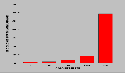
Counting the number of B. larvae colonies per plate can provide a better indication of hives likely to contain diseased larvae. The table at the end of this section shows that the greater the number of B. larvae colonies per plate, the greater the probability that the colony already shows visual signs of American foulbrood. (Note: this study used 60 Ál of sample material applied per plate, rather than the 20 Ál per plate recommended in this manual).
If a colony tests positive, every brood frame should be thoroughly checked as soon as possible. If diseased larvae are found, the colony must be burnt. If no diseased larvae are found, the colony should be marked and quarantined for one full year. At the end of that period, another complete brood check should be carried out together with a culture test using adult bees. If a hive tests positive again, but does not show visual signs of the disease, it should be left in quarantine for another full year.
During the quarantine period, no hive parts should be removed from the colony or placed on any other colony. It might be worth moving positive colonies to a separate site were they can be inspected more often.

Glassware can be sterilized in a household pressure cooker.
To check that the sterilizing method is working, place some control AFB solution (see Appendix 3) in a bottle and sterilize as above. Plate the material to see if the spores have been killed.
The following is a list of suppliers and prices for lab materials referred to in this manual. There are a number of sources for most of these items. However, Labsupply Pierce (see below) is recommended because they will fill small orders.
Some of the materials (eg. hydrogen peroxide) are supplied in greater amounts and stronger concentrations than what you will probably require. In such cases you may find that other outlets may be able to supply smaller quantities and lower concentrations (eg. chemist shops). You may also be able to replace some of the equipment with cheaper, household items. You should consider carefully, however, the item's requirements before purchasing alternatives.
1) Labsupply Pierce
P.O.Box 34234
Birkenhead
AUCKLAND 10
Ph. 0800 734 100
| Bottle (Schott) | 500 ml | $8.54 |
| Bottle (Schott) | 1 litre | 12.11 |
| Bottle (Universal) | 28 ml | 1.15 |
| Forceps, blunt stainless steel | 11.5 cm | 2.75 |
| Measuring cylinder, graduated glass | 10 ml | 5.47 |
| Thermometer, red spirit, general purpose 300mm, 75mm immersion, |
-10 to 110oC | 8.50 |
| Burner, spirit | 100 ml | 14.52 |
| Hydrogen peroxide | 100 volumes (30%) 2 litre | 24.56 |
| Thiamine Hydrochloride (Vitamin B1) | 25 g | 47.50 |
| Petri dish, disposable, (per 500) | 70.00 | |
| Pasteur pipettes, glass (per250) | 150 mm | 18.55 |
| Automatic pipette, non-adjustable | 60 Ál | 214.00 |
| Pipette tips (per 1000) | 60 Ál | 84.52 |
2) NDA Labware
Auckland
Ph. 09 525 1030
| Wire loops (per 100) | 10 Ál | 10.00 |
3) Fort Richards Lab
PO Box 22192
Auckland
Ph. 09 276 5569
| Difco Brain Heart Infusion (BHI), agar | 454 g | 139.30 |
4) Convex Plastic Ltd
Hamilton
Ph. 07 847 5133
| Plastic bags (per 1,000) | 15cm x 13cm | 11.10 |
A solution containing B. larvae spores can be very useful for infecting control plates. Incubated control plates let you know if a) you are preparing and infecting your plates properly, b) your incubator temperature is set correctly, and c) excess heat during the processing of samples is destroying viable B. larvae spores.
To make the solution:
Too Few Colonies - if only a few B. larvae colonies grow on the plates, the solution is not concentrated enough. You will need to make up a more concentrated solution using the remainder of the original 10 ml solution.
Too Many Colonies - if there is so much growth on the plates that you cannot see distinct colonies, dilute the solution with another 50 ml of sterile water.
Storage - When the solution is at the required concentration it should be marked clearly and kept in a refrigerator. In this form, spores should remain viable almost indefinitely.
Expensive, manufactured incubators are not necessary to grow out bacterial plates. A perfectly adequate incubator can be constructed from simple materials which can either be scrounged or purchased from local suppliers. The important thing is to ensure that the required temperature is kept relatively constant and that the heat does not "layer" in the incubator unit.
A discarded refrigerator can easily be made into a good incubator. Small, motel-size refrigerators are excellent for this purpose. If a refrigerator is not available, build a box from plywood large enough to take the number of plates you wish to incubate at one time. The box should be made with both inner and outer walls and should be lagged with household insulation (batts). The door should fit tightly and have a rubber sealing gasket.
A variety of heat sources can be used, including seed starter mats (available from garden centres), small fan heaters, and 40 watt lights. Two 40 watt light bulbs wired in parallel will provide sufficient heat for a standard size refrigerator.
Commonly available thermostats which can be used include egg incubator thermostats (available from farm supply stores) and aquarium thermostats (available from pet stores). You may need to consult an electrician to properly wire the thermostat to the heating source.
Attach the thermostat midway up the side way of the incubator, rather than at the top or bottom. This will avoid false readings which could affect the growth of your cultures. Larger incubators (full refrigerator size) may also need a small fan to ensure that heat is properly circulated. Car fans are ideal for this purpose, but must be wired through a transformer if they are not 240v. The fan should be wired to run constantly, even when the thermostat has turned the heater off.
To check whether the incubator is working properly, infect six or plates with control solution (see "Control Plates" in the Infecting Plates section). Place the plates in different positions in the incubator (top, bottom, back, front). If the incubator is producing the correct temperature and the air inside is being properly circulated, all plates should produce similar numbers of B. larvae colonies.
Home NZ Bkpg Bee Diseases Organisation Information Contacts
Email to Nick Wallingford, webmaster of the site...